


Factors Affecting Wound Healing
Wound healing, as a normal biological process in the human body, is achieved through four precisely and highly programmed phases: hemostasis, inflammation, proliferation, and remodeling. For a wound to heal successfully, all four phases must occur in the proper sequence and time frame. Many factors can interfere with one or more phases of this process, thus causing improper or impaired wound healing. This article reviews the recent literature on the most significant factors that affect cutaneous wound healing and the potential cellular and/or molecular mechanisms involved. The factors discussed include oxygenation, infection, age and sex hormones, stress, diabetes, obesity, medications, alcoholism, smoking, and nutrition. A better understanding of the influence of these factors on repair may lead to therapeutics that improve wound healing and resolve impaired wounds.
INTRODUCTION
The wound-healing process consists of four highly integrated and overlapping phases: hemostasis, inflammation, proliferation, and tissue remodeling or resolution (Gosain and DiPietro, 2004). These phases and their biophysiological functions must occur in the proper sequence, at a specific time, and continue for a specific duration at an optimal intensity (Table 1; Mathieu et al., 2006). There are many factors that can affect wound healing which interfere with one or more phases in this process, thus causing improper or impaired tissue repair.
Table 1.
Normal Wound-healing Process
| Phase | Cellular and Bio-physiologic Events |
|---|---|
| Hemostasis |
|
| Inflammation |
|
| Proliferation |
|
| Remodeling |
|
ECM, extracellular matrix.
Wounds that exhibit impaired healing, including delayed acute wounds and chronic wounds, generally have failed to progress through the normal stages of healing. Such wounds frequently enter a state of pathologic inflammation due to a postponed, incomplete, or uncoordinated healing process. Most chronic wounds are ulcers that are associated with ischemia, diabetes mellitus, venous stasis disease, or pressure. Non-healing wounds affect about 3 to 6 million people in the United States, with persons 65 years and older accounting for 85% of these events. Non-healing wounds result in enormous health care expenditures, with the total cost estimated at more than $3 billion per year (Mathieu et al., 2006; Menke et al., 2007).
Laboratory investigations and clinical studies have yielded a wealth of information about both normal and impaired wound healing. More recently, a great deal of research has been directed at understanding the critical factors that influence poorly healing wounds. While much remains to be learned, these studies may lead to therapeutics that will promote proper tissue repair and improve impaired wound healing. This review will discuss the many different factors that affect cutaneous wound healing and the potential cellular and molecular mechanisms involved.
THE WOUND-HEALING PROCESS
Wound healing is a dynamic process consisting of four continuous, overlapping, and precisely programmed phases. The events of each phase must happen in a precise and regulated manner. Interruptions, aberrancies, or prolongation in the process can lead to delayed wound healing or a non-healing chronic wound.
In adult humans, optimal wound healing involves the following the events: (1) rapid hemostasis; (2) appropriate inflammation; (3) mesenchymal cell differentiation, proliferation, and migration to the wound site; (4) suitable angiogenesis; (5) prompt re-epithelialization (re-growth of epithelial tissue over the wound surface); and (6) proper synthesis, cross-linking, and alignment of collagen to provide strength to the healing tissue (Gosain and DiPietro, 2004; Mathieu et al., 2006). The first phase of hemostasis begins immediately after wounding, with vascular constriction and fibrin clot formation. The clot and surrounding wound tissue release pro-inflammatory cytokines and growth factors such as transforming growth factor (TGF)-β, platelet-derived growth factor (PDGF), fibroblast growth factor (FGF), and epidermal growth factor (EGF). Once bleeding is controlled, inflammatory cells migrate into the wound (chemotaxis) and promote the inflammatory phase, which is characterized by the sequential infiltration of neutrophils, macrophages, and lymphocytes (Gosain and DiPietro, 2004; Broughton et al., 2006; Campos et al., 2008). A critical function of neutrophils is the clearance of invading microbes and cellular debris in the wound area, although these cells also produce substances such as proteases and reactive oxygen species (ROS), which cause some additional bystander damage.
Macrophages play multiple roles in wound healing. In the early wound, macrophages release cytokines that promote the inflammatory response by recruiting and activating additional leukocytes. Macrophages are also responsible for inducing and clearing apoptotic cells (including neutrophils), thus paving the way for the resolution of inflammation. As macrophages clear these apoptotic cells, they undergo a phenotypic transition to a reparative state that stimulates keratinocytes, fibroblasts, and angiogenesis to promote tissue regeneration (Meszaros et al., 2000; Mosser and Edwards, 2008). In this way, macrophages promote the transition to the proliferative phase of healing.
T-lymphocytes migrate into wounds following the inflammatory cells and macrophages, and peak during the late-proliferative/early-remodeling phase. The role of T-lymphocytes is not completely understood and is a current area of intensive investigation. Several studies suggest that delayed T-cell infiltration along with decreased T-cell concentration in the wound site is associated with impaired wound healing, while others have reported that CD 4+ cells (T-helper cells) have a positive role in wound healing and CD8+ cells (T-suppressor-cytotoxic cells) play an inhibitory role in wound healing (Swift et al., 2001; Park and Barbul, 2004). Interestingly, recent studies in mice deficient in both T- and B-cells have shown that scar formation is diminished in the absence of lymphocytes (Gawronska-Kozak et al., 2006). In addition, skin gamma-delta T-cells regulate many aspects of wound healing, including maintaining tissue integrity, defending against pathogens, and regulating inflammation. These cells are also called dendritic epidermal T-cells (DETC), due to their unique dendritic morphology. DETC are activated by stressed, damaged, or transformed keratinocytes and produce fibroblast growth factor 7 (FGF-7), keratinocyte growth factors, and insulin-like growth factor-1, to support keratinocyte proliferation and cell survival. DETC also generate chemokines and cytokines that contribute to the initiation and regulation of the inflammatory response during wound healing. While cross-talk between skin gamma-delta T-cells and keratinocytes contributes to the maintenance of normal skin and wound healing, mice lacking or defective in skin gamma-delta T-cells show a delay in wound closure and a decrease in the proliferation of keratinocytes at the wound site (Jameson and Havran, 2007; Mills et al., 2008).
The proliferative phase generally follows and overlaps with the inflammatory phase, and is characterized by epithelial proliferation and migration over the provisional matrix within the wound (re-epithelialization). In the reparative dermis, fibroblasts and endothelial cells are the most prominent cell types present and support capillary growth, collagen formation, and the formation of granulation tissue at the site of injury. Within the wound bed, fibroblasts produce collagen as well as glycosaminoglycans and proteoglycans, which are major components of the extracellular matrix (ECM). Following robust proliferation and ECM synthesis, wound healing enters the final remodeling phase, which can last for years. In this phase, regression of many of the newly formed capillaries occurs, so that vascular density of the wound returns to normal. One critical feature of the remodeling phase is ECM remodeling to an architecture that approaches that of the normal tissue. The wound also undergoes physical contraction throughout the entire wound-healing process, which is believed to be mediated by contractile fibroblasts (myofibroblasts) that appear in the wound (Gosain and DiPietro, 2004; Campos et al., 2008).
The role of stem cells (SC) in cutaneous wound healing and tissue regeneration is a topic of increasing research attention, with a focus on the role of adult stem cells such as epidermal stem cells and bone-marrow (BM)-derived cells (BMDCs). Epidermal stem cells reside in the bulge area of hair follicles and in the basal layer of the epidermis and give rise to the keratinocytes that migrate andre-epithelialize wounds. Normal skin is also a target organ for BMDCs. Two main stem cell populations are present in the bone marrow: hematopoietic SC (HSC) and mesenchymal SC (MSC). BM-MSCs are able to differentiate into a variety of cell types, including adipocytes, osteoblasts, chondrocytes, fibroblasts, and keratinocytes (Cha and Falanga, 2007; Rea et al., 2009). Endothelial progenitor cells (EPCs) derived from the HSC lineage are key cells that contribute to neovascularization. Both BM-MSCs and EPCs are involved in the cutaneous wound-healing process. Wound-induced hypoxia triggers the mobilization of bone marrow EPCs to the circulation, playing a significant role in the process of neovascularization (Wu et al., 2007; Liu and Velazquez, 2008; Rea et al., 2009).
Several different cell types are involved in the wound-healing process, and, as described above, the cellular activities of any particular cell type may also vary during different stages of repair. The complexity and coordination of the healing process are major hurdles to therapeutic approaches, since any therapeutic must effectively be sequenced to the appropriate stage.
FACTORS AFFECTING WOUND HEALING
Multiple factors can lead to impaired wound healing. In general terms, the factors that influence repair can be categorized into local and systemic. Local factors are those that directly influence the characteristics of the wound itself, while systemic factors are the overall health or disease state of the individual that affect his or her ability to heal (Table 2). Many of these factors are related, and the systemic factors act through the local effects affecting wound healing.
Table 2.
Factors Affecting Wound Healing
| Local Factors | Systemic Factors |
|---|---|
|
|
Local Factors That Influence Healing
Oxygenation
Oxygen is important for cell metabolism, especially energy production by means of ATP, and is critical for nearly all wound-healing processes. It prevents wounds from infection, induces angiogenesis, increases keratinocyte differentiation, migration, and re-epithelialization, enhances fibroblast proliferation and collagen synthesis, and promotes wound contraction (Bishop, 2008; Rodriguez et al., 2008). In addition, the level of superoxide production (a key factor for oxidative killing pathogens) by polymorphonuclear leukocytes is critically dependent on oxygen levels.
Due to vascular disruption and high oxygen consumption by metabolically active cells, the microenvironment of the early wound is depleted of oxygen and is quite hypoxic. Several systemic conditions, including advancing age and diabetes, can create impaired vascular flow, thus setting the stage for poor tissue oxygenation. In the context of healing, this overlay of poor perfusion creates a hypoxic wound. Chronic wounds are notably hypoxic; tissue oxygen tensions have been measured transcutaneously in chronic wounds from 5 to 20 mm Hg, in contrast to control tissue values of 30 to 50 mm Hg (Tandara and Mustoe, 2004).
In wounds where oxygenation is not restored, healing is impaired. Temporary hypoxia after injury triggers wound healing, but prolonged or chronic hypoxia delays wound healing (Bishop, 2008; Rodriguez et al., 2008). In acute wounds, hypoxia serves as a signal that stimulates many aspects of the wound-healing process. Hypoxia can induce cytokine and growth factor production from macrophages, keratinocytes, and fibroblasts. Cytokines that are produced in response to hypoxia include PDGF, TGF-β, VEGF, tumor necrosis factor-α (TNF-α), and endothelin-1, and are crucial promoters of cell proliferation, migration and chemotaxis, and angiogenesis in wound healing (Rodriguez et al., 2008).
In normally healing wounds, ROS such as hydrogen peroxide (H2O2) and superoxide (O2) are thought to act as cellular messengers to stimulate key processes associated with wound healing, including cell motility, cytokine action (including PDGF signal transduction), and angiogenesis. Both hypoxia and hyperoxia increase ROS production, but an increased level of ROS transcends the beneficial effect and causes additional tissue damage (Rodriguez et al., 2008).
In summary, the proper oxygen level is crucial for optimum wound healing. Hypoxia stimulates wound healing such as the release of growth factors and angiogenesis, while oxygen is needed to sustain the healing process (Bishop, 2008). One therapeutic option that can sometimes overcome the influence of tissue hypoxia is hyperbaric oxygen therapy (HBOT; Rodriguez et al., 2008). While HBOT can be an effective treatment for hypoxic wounds, its availability is limited.
Infections
Once skin is injured, micro-organisms that are normally sequestered at the skin surface obtain access to the underlying tissues. The state of infection and replication status of the micro-organisms determine whether the wound is classified as having contamination, colonization, local infection/critical colonization, and/or spreading invasive infection. Contamination is the presence of non-replicating organisms on a wound, while colonization is defined as the presence of replicating micro-organisms on the wound without tissue damage. Local infection/critical colonization is an intermediate stage, with micro-organism replication and the beginning of local tissue responses. Invasive infection is defined as the presence of replicating organisms within a wound with subsequent host injury (Edwards and Harding, 2004).
Inflammation is a normal part of the wound-healing process, and is important to the removal of contaminating micro-organisms. In the absence of effective decontamination, however, inflammation may be prolonged, since microbial clearance is incomplete. Both bacteria and endotoxins can lead to the prolonged elevation of pro-inflammatory cytokines such as interleukin-1 (IL-1) and TNF-α and elongate the inflammatory phase. If this continues, the wound may enter a chronic state and fail to heal. This prolonged inflammation also leads to an increased level of matrix metalloproteases (MMPs), a family of proteases that can degrade the ECM. In tandem with the increased protease content, a decreased level of the naturally occurring protease inhibitors occurs. This shift in protease balance can cause growth factors that appears in chronic wounds to be rapidly degraded (Edwards and Harding, 2004; Menke et al., 2007). Similar to other infective processes, the bacteria in infected wounds occur in the form of biofilms, which are complex communities of aggregated bacteria embedded in a self-secreted extracellular polysaccharide matrix (EPS; Edwards and Harding, 2004). Mature biofilms develop protected microenvironments and are more resistant to conventional antibiotic treatment. Staphylococcus aureus (S. aureus), Pseudomonas aeruginosa (P. aeruginosa), and β-hemolytic streptococci are common bacteria in infected and clinically non-infected wounds (Edwards and Harding, 2004; Davis et al., 2008).
P. aeruginosa and Staphylococcus appear to play an important role in bacterial infection in wounds. Many chronic ulcers probably do not heal because of the presence of biofilms containing P. aeruginosa, thus shielding the bacteria from the phagocytic activity of invading polymorphonuclear neutrophils (PMNs). This mechanism may explain the failure of antibiotics as a remedy for chronic wounds (Bjarnsholt et al., 2008).
Systemic Factors That Influence Healing
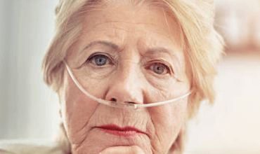
Age
The elderly population (people over 60 years of age) is growing faster than any other age group (World Health Organization [WHO, www.who.int/topics/ageing]), and increased age is a major risk factor for impaired wound healing. Many clinical and animal studies at the cellular and molecular level have examined age-related changes and delays in wound healing. It is commonly recognized that, in healthy older adults, the effect of aging causes a temporal delay in wound healing, but not an actual impairment in terms of the quality of healing (Gosain and DiPietro, 2004; Keylock et al., 2008). Delayed wound healing in the aged is associated with an altered inflammatory response, such as delayed T-cell infiltration into the wound area with alterations in chemokine production and reduced macrophage phagocytic capacity (Swift et al., 2001). Delayed re-epithelialization, collagen synthesis, and angiogenesis have also been observed in aged mice as compared with young mice (Swift et al., 1999). Overall, there are global differences in wound healing between young and aged individuals. A review of the age-related changes in healing capacity demonstrates that every phase of healing undergoes characteristic age-related changes, including enhanced platelet aggregation, increased secretion of inflammatory mediators, delayed infiltration of macrophages and lymphocytes, impaired macrophage function, decreased secretion of growth factors, delayed re-epithelialization, delayed angiogenesis and collagen deposition, reduced collagen turnover and remodeling, and decreased wound strength (Gosain and DiPietro, 2004).
Several treatments to reduce the age-related impairment of healing have been studied. Interestingly, exercise has been reported to improve cutaneous wound healing in older adults as well as aged mice, and the improvement is associated with decreased levels of pro-inflammatory cytokines in the wound tissue. The improved healing response may be due to an exercise-induced anti-inflammatory response in the wound (Emery et al., 2005; Keylock et al., 2008).
Sex Hormones in Aged Individuals
Sex hormones play a role in age-related wound-healing deficits. Compared with aged females, aged males have been shown to have delayed healing of acute wounds. A partial explanation for this is that the female estrogens (estrone and 17β-estradiol), male androgens (testosterone and 5α-dihydrotestosterone, DHT), and their steroid precursor dehydroepiandrosterone (DHEA) appear to have significant effects on the wound-healing process (Gilliver et al., 2007). It was recently found that the differences in gene expression between elderly male and young human wounds are almost exclusively estrogen-regulated (Hardman and Ashcroft, 2008). Estrogen affects wound healing by regulating a variety of genes associated with regeneration, matrix production, protease inhibition, epidermal function, and the genes primarily associated with inflammation (Hardman and Ashcroft, 2008). Studies indicate that estrogen can improve the age-related impairment in healing in both men and women, while androgens regulate cutaneous wound healing negatively (Gilliver et al., 2007).
Stress
Stress has a great impact on human health and social behavior. Many diseases—such as cardiovascular disease, cancer, compromised wound healing, and diabetes—are associated with stress. Numerous studies have confirmed that stress-induced disruption of neuroendocrine immune equilibrium is consequential to health (Glaser and Kiecolt-Glaser, 2005; Vileikyte, 2007). The pathophysiology of stress results in the deregulation of the immune system, mediated primarily through the hypothalamic-pituitary-adrenal (HPA) and sympathetic-adrenal medullary axes or sympathetic nervous system (SNS; Godbout and Glaser, 2006; Boyapati and Wang, 2007).
Studies in both humans and animals have demonstrated that psychological stress causes a substantial delay in wound healing. Caregivers of persons with Alzheimer’s and students undergoing academic stress during examinations demonstrated delayed wound healing (Kiecolt-Glaser et al., 1995; Marucha et al., 1998). The hypothalamic-pituitary-adrenal and the sympathetic-adrenal medullary axes regulate the release of pituitary and adrenal hormones. These hormones include the adrenocorticotrophic hormones, cortisol and prolactin, and catecholamines (epinephrine and norepinephrine). Stress up-regulates glucocorticoids (GCs) and reduces the levels of the pro-inflammatory cytokines IL-1β, IL-6, and TNF-α at the wound site. Stress also reduces the expression of IL-1α and IL-8 at wound sites—both chemoattractants that are necessary for the initial inflammatory phase of wound healing (Godbout and Glaser, 2006; Boyapati and Wang, 2007). Furthermore, GCs influence immune cells by suppressing differentiation and proliferation, regulating gene transcription, and reducing expression of cell adhesion molecules that are involved in immune cell trafficking (Sternberg, 2006). The GC cortisol functions as an anti-inflammatory agent and modulates the Th1-mediated immune responses that are essential for the initial phase of healing. Thus, psychological stress impairs normal cell-mediated immunity at the wound site, causing a significant delay in the healing process (Godbout and Glaser, 2006).
Stressors can lead to negative emotional states, such as anxiety and depression, which may in turn have an impact on physiologic processes and/or behavioral patterns that influence health outcomes. In addition to the direct influences of anxiety and depression on endocrine and immune function, stressed individuals are more likely to have unhealthy habits, which include poor sleep patterns, inadequate nutrition, less exercise, and a greater propensity for abuse of alcohol, cigarettes, and other drugs. All of these factors may come into play in negatively modulating the healing response. The effects of stress on wound healing are summarized in Fig. 1.

The effects of stress on wound healing. Stress-impaired wound healing is mediated primarily through the hypothalamic-pituitary-adrenal, sympathetic-adrenal medullary axes, and psychological-response-induced unhealthy behaviors.
Diabetes
Diabetes affects hundreds of millions of people worldwide. Diabetic individuals exhibit a documented impairment in the healing of acute wounds. Moreover, this population is prone to develop chronic non-healing diabetic foot ulcers (DFUs), which are estimated to occur in 15% of all persons with diabetes. DFUs are a serious complication of diabetes, and precede 84% of all diabetes-related lower leg amputations (Brem and Tomic-Canic, 2007). The impaired healing of both DFUs and acute cutaneous wounds in persons with diabetes involves multiple complex pathophysiological mechanisms. DFUs, like venous stasis disease and pressure-related chronic non-healing wounds, are always accompanied by hypoxia (Tandara and Mustoe, 2004). A situation of prolonged hypoxia, which may be derived from both insufficient perfusion and insufficient angiogenesis, is detrimental for wound healing. Hypoxia can amplify the early inflammatory response, thereby prolonging injury by increasing the levels of oxygen radicals (Mathieu et al., 2006; Woo et al., 2007). Hyperglycemia can also add to the oxidative stress when the production of ROS exceeds the anti-oxidant capacity (Vincent et al., 2004). The formation of advanced glycation end-products (AGEs) under hyperglycemia and the interaction with their receptors (RAGE) are associated with impaired wound healing in diabetic mice as well (Huijberts et al., 2008). High levels of metalloproteases are a feature of diabetic foot ulcers, and the MMP levels in chronic wound fluid are almost 60 times higher than those in acute wounds. This increased protease activity supports tissue destruction and inhibits normal repair processes (Woo et al., 2007; Sibbald and Woo, 2008).
Several dysregulated cellular functions are involved in diabetic wounds, such as defective T-cell immunity, defects in leukocyte chemotaxis, phagocytosis, and bactericidal capacity, and dysfunctions of fibroblasts and epidermal cells. These defects are responsible for inadequate bacterial clearance and delayed or impaired repair in individuals with diabetes (Loots et al., 1998; Sibbald and Woo, 2008).
As mentioned above, hypoxia contributes to the compromised healing of DFUs, and diabetic wounds exhibit inadequate angiogenesis. Several studies that have investigated the mechanisms behind the decreased restoration of vasculature in diabetic wounds have implied that EPC mobilization and homing are impaired, and that the level of VEGF, the primary pro-angiogenic factor in wounds, is decreased in the diabetic state (Brem and Tomic-Canic, 2007; Gallagher et al., 2007; Quattrini et al., 2008). Stem-cell-based therapies aimed at inducing EPCs or BM-MSCs have shown a promising outcome in diabetic non-healing wounds, both in animals and in clinical trials (Wu et al., 2007; Liu and Velazquez, 2008; Rea et al., 2009). In animal studies, therapeutic restoration of VEGF has been shown to improve repair outcomes significantly (Kirchner et al., 2003; Galiano et al., 2004).
The neuropathy that occurs in diabetic individuals probably also contributes to impaired wound healing. Neuropeptides such as nerve growth factor, substance P, and calcitonin gene-related peptide are relevant to wound healing, because they promote cell chemotaxis, induce growth factor production, and stimulate the proliferation of cells. A decrease in neuropeptides has been associated with DFU formation. In addition, sensory nerves play a role in modulating immune defense mechanisms, with denervated skin exhibiting reduced leukocyte infiltration (Galkowska et al., 2006; Sibbald and Woo, 2008).
In summary, the impaired healing that occurs in individuals with diabetes involves hypoxia, dysfunction in fibroblasts and epidermal cells, impaired angiogenesis and neovascularization, high levels of metalloproteases, damage from ROS and AGEs, decreased host immune resistance, and neuropathy. The influence of these factors on wound healing is summarized in Fig. 2.

The potential effects of diabetes on wound healing. MMPs, matrix metalloproteases; ROS, reactive oxygen species; AGEs, advanced glycation end-products.
Medications
Many medications, such as those which interfere with clot formation or platelet function, or inflammatory responses and cell proliferation have the capacity to affect wound healing. Here we review only the commonly used medications that have a significant impact on healing, including glucocorticoid steroids, non-steroidal anti-inflammatory drugs, and chemotherapeutic drugs.
Glucocorticoid Steroids
Systemic glucocorticoids (GC), which are frequently used as anti-inflammatory agents, are well-known to inhibit wound repair via global anti-inflammatory effects and suppression of cellular wound responses, including fibroblast proliferation and collagen synthesis. Systemic steroids cause wounds to heal with incomplete granulation tissue and reduced wound contraction (Franz et al., 2007). Glucocorticoids also inhibit production of hypoxia-inducible factor-1 (HIF-1), a key transcriptional factor in healing wounds (Wagner et al., 2008). Beyond effects on repair itself, systemic corticosteroids may increase the risk of wound infection. While systemic corticosteroids inhibit wound repair, topical application produces quite different effects. Topical low-dosage corticosteroid treatment of chronic wounds has been found to accelerate wound healing, reduce pain and exudate, and suppress hypergranulation tissue formation in 79% of cases. While these positive effects are striking, careful monitoring is necessary to avoid a potential increased risk of infection with prolonged use (Hofman et al., 2007).
Non-steroidal Anti-inflammatory Drugs
Non-steroidal anti-inflammatory drugs (NSAIDs) such as ibuprofen are widely used for the treatment of inflammation and rheumatoid arthritis and for pain management. Low-dosage aspirin, due to its anti-platelet function, is commonly used as a preventive therapeutic for cardiovascular disease, but not as an anti-inflammatory drug (Pieringer et al., 2007). There are few data to suggest that short-term NSAIDs have a negative impact on healing. However, the question of whether long-term NSAIDs interfere with wound healing remains open. In animal models, systemic use of ibuprofen has demonstrated an anti-proliferative effect on wound healing, resulting in decreased numbers of fibroblasts, weakened breaking strength, reduced wound contraction, delayed epithelialization (Dong et al., 1993; Dvivedi et al., 1997; Krischak et al., 2007), and impaired angiogenesis (Jones et al., 1999). The effects of low-dose aspirin on healing are not completely clear. Clinical recommendations suggest that, to avoid anti-platelet effects, individuals should discontinue NSAIDs for a time period equal to 4 to 5 times the half-life of drugs before surgery. Thus, the majority of surgical patients do not have significant NSAID activity at the time of wound repair. The exception may be those cardiac patients who must be maintained on low-dose aspirin due to severe risk of cardiovascular events (Pieringer et al., 2007). In terms of the topical application of NSAIDs on the surfaces of chronic wounds, the local use of ibuprofen-foam provides moist wound healing, reduces persistent and temporary wound pain, and benefits chronic venous leg ulcer healing (Price et al., 2007).
Chemotherapeutic Drugs
Most chemotherapeutic drugs are designed to inhibit cellular metabolism, rapid cell division, and angiogenesis and thus inhibit many of the pathways that are critical to appropriate wound repair. These medications inhibit DNA, RNA, or protein synthesis, resulting in decreased fibroplasia and neovascularization of wounds (Waldron and Zimmerman-Pope, 2003; Franz et al., 2007). Chemotherapeutic drugs delay cell migration into the wound, decrease early wound matrix formation, lower collagen production, impair proliferation of fibroblasts, and inhibit contraction of wounds (Franz et al., 2007). In addition, these agents weaken the immune functions of the patients, and thereby impede the inflammatory phase of healing and increase the risk of wound infection. Chemotherapy induces neutropenia, anemia, and thrombocytopenia, thus leaving wounds vulnerable to infection, causing less oxygen delivery to the wound, and also making patients vulnerable to excessive bleeding at the wound site.
Impaired wound healing due to chemotherapeutic drugs such as adriamycin is most common when the drugs are administered pre-operatively or within 3 weeks post-operatively (Lawrence et al., 1986). Additionally, low post-operative albumin levels, low post-operative hemoglobin, advanced stage of disease, and electrocautery use have all been reported as risk factors for the development of wound complications (Kolb et al., 1992).
A newer generation of tumor chemotherapeutics is the angiogenesis inhibitors, such as bevacizumab, which is an antibody fragment that neutralizes VEGF. These therapies work in conjunction with traditional chemotherapeutics to limit the blood supply to tumors, reducing their ability to grow. Wound-healing complications, including an increase in wound dehiscence, have been described in patients on angiogenesis inhibitors (Lemmens et al., 2008). A caveat is that most patients on angiogenesis inhibitors are also on traditional chemotherapeutics, making it difficult to sort out whether angiogenesis inhibitors alone would perturb repair (Scappaticci et al., 2005; Scott, 2007). Nevertheless, current recommendations include discontinuation of angiogenesis inhibitors well in advance of any surgical procedures.
Obesity
The prevalence of obesity continues to increase among adults, children, and adolescents in the United States, with more than 30% of adults and 15% of children and adolescents classified as obese in a recent survey (Centers for Disease Control and Prevention, CDC). Obesity is well-known to increase the risk of many diseases and health conditions, which include coronary heart disease, type 2 diabetes, cancer, hypertension, dyslipidemia, stroke, sleep apnea, respiratory problems, and impaired wound healing. Obese individuals frequently face wound complications, including skin wound infection, dehiscence, hematoma and seroma formation, pressure ulcers, and venous ulcers (Wilson and Clark, 2004; www.cdc.gov/nccdphp/dnpa/obesity). An increased frequency of wound complications has been reported for obese individuals undergoing both bariatric and non-bariatric operations (Anaya and Dellinger, 2006; Greco et al., 2008; Momeni et al., 2009). In particular, a higher rate of surgical site infection occurs in obese patients. Many of these complications may be a result of a relative hypoperfusion and ischemia that occurs in subcutaneous adipose tissue. This situation may be caused by a decreased delivery of antibiotics as well. In surgical wounds, the increased tension on the wound edges that is frequently seen in obese patients also contributes to wound dehiscence. Wound tension increases tissue pressure, reducing microperfusion and the availability of oxygen to the wound (Wilson and Clark, 2004; Anaya and Dellinger, 2006).
The increase in pressure ulcers or pressure-related injuries in obese individuals is also influenced by hypovascularity, since poor perfusion makes tissue more susceptible to this type of injury. In addition, the difficulty or inability of obese individuals to reposition themselves further increases the risk of pressure-related injuries. Moreover, skin folds harbor micro-organisms that thrive in moist areas and contribute to infection and tissue breakdown. The friction caused by skin-on-skin contact invites ulceration. Together, these factors predispose obese individuals to the development of impaired wound healing (Wilson and Clark, 2004; Anaya and Dellinger, 2006; Greco et al., 2008).
In addition to local conditions, systemic factors also play an important role in impaired wound healing and wound complications in obese patients. Obesity can be connected to stress, anxiety, and depression, all situations which can cause an impaired immune response (Wilson and Clark, 2004).
The function of adipose tissue used to be considered as primarily caloric storage. However, more recent findings have documented that adipose tissue secretes a large variety of bioactive substances that are collectively named adipokines. Both adipocytes themselves as well as macrophages inside the adipose tissue are known to produce bioactive molecules including cytokines, chemokines, and hormone-like factors such as leptin, adiponectin, and resistin. Adipokines have a profound impact on the immune and inflammatory response (Juge-Aubry et al., 2005; Calabro and Yeh, 2007; Wozniak et al., 2009). The negative influence of adipokines on the systemic immune response seems likely to influence the healing process, although direct proof for this is lacking. Impaired peripheral blood mononuclear cell function, decreased lymphocyte proliferation, and altered peripheral cytokine levels have been reported in obesity. Importantly, though, many of the obesity-related changes in peripheral immune function are improved by weight loss (Nieman et al., 1999; Fontana et al., 2007; de Mello et al., 2008). The factors related to wound impairment in obesity are summarized in Table 3.
Table 3.
Summary of Factors Related to Wound Impairment in Obesity
| Local Wound Conditions | Associated Diseases and Conditions | Factors Altering Immune and Inflammatory Responses |
|---|---|---|
|
|
|
MCP, monocyte chemoattractant protein-1; IP-10 interferon-gamma-inducible protein 10.
Alcohol Consumption
Clinical evidence and animal experiments have shown that exposure to alcohol impairs wound healing and increases the incidence of infection (Gentilello et al., 1993; Szabo and Mandrekar, 2009). The effect of alcohol on repair is quite clinically relevant, since over half of all emergency room trauma cases involve either acute or chronic alcohol exposure (Rivara et al., 1993; Madan et al., 1999). Alcohol exposure diminishes host resistance, and ethanol intoxication at the time of injury is a risk factor for increased susceptibility to infection in the wound (Choudhry and Chaudry, 2006). Studies have demonstrated profound effects of alcohol on host-defense mechanisms, although the precise effects are dependent upon the pattern of alcohol exposure (i.e., chronic vs. acute alcohol exposure, amount consumed, duration of consumption, time from alcohol exposure, and alcohol withdrawal). A recent review on alcohol-induced alterations on host defense after traumatic injury suggested that, in general, short-term acute alcohol exposure results in suppressed pro-inflammatory cytokine release in response to an inflammatory challenge. The higher rate of post-injury infection correlates with decreased neutrophil recruitment and phagocytic function in acute alcohol exposure (Greiffenstein and Molina, 2008).
Beyond the increased incidence of infection, exposure to ethanol also seems to influence the proliferative phase of healing. In murine models, exposure to a single dose of alcohol that caused a blood alcohol level of 100 mg/dL (just above the legal limit in most states in the US) perturbed re-epithelialization, angiogenesis, collagen production, and wound closure (Radek et al., 2005, 2007, 2008; Fitzgerald et al., 2007). The most significant impairment seems to be in wound angiogenesis, which is reduced by up to 61% following a single ethanol exposure. This decrease in angiogenic capacity involves both decreased expression of VEGF receptors and reduced nuclear expression of HIF-1alpha in endothelial cells (Radek et al., 2005, 2008). The ethanol-mediated decrease in wound vascularity causes increased wound hypoxia and oxidative stress (Radek et al., 2008). Connective tissue restoration is also influenced by acute ethanol exposure, and results in decreased collagen production and alterations in the protease balance at the wound site (Radek et al., 2007). In summary, acute ethanol exposure can lead to impaired wound healing by impairing the early inflammatory response, inhibiting wound closure, angiogenesis, and collagen production, and altering the protease balance at the wound site.
As mentioned previously, the host response to chronic alcohol exposure appears to be different from that of acute alcohol exposure. Analysis of clinical data indicates that chronic alcohol exposure causes impaired wound healing and enhanced host susceptibility to infections, but the detailed mechanisms that explain this effect need more investigation.
Smoking
It is well-known that smoking increases the risk of heart and vascular disease, stroke, chronic lung disease, and many kinds of cancers. Similarly, the negative effects of smoking on wound-healing outcomes have been known for a long time (Siana et al., 1989; Jensen et al., 1991; Ahn et al., 2008). Post-operatively, patients who smoke show a delay in wound healing and an increase in a variety of complications such as infection, wound rupture, anastomotic leakage, wound and flap necrosis, epidermolysis, and a decrease in the tensile strength of wounds (Chan et al., 2006; Ahn et al., 2008). In the realm of oral surgery, impaired healing in smokers has been noticed both in routine oral surgery and in the placement of dental implants (Levin and Schwartz-Arad, 2005; Balaji, 2008). Cosmetic outcomes also appear to be worse in smokers, and plastic and reconstructive surgeons are often reluctant to perform cosmetic surgeries on individuals who refuse to quit smoking (Siana et al., 1989; Bennett, 1991). Approximately over 4000 substances in tobacco smoke have been identified, and some have been shown to have a negative impact on healing (Ahn et al., 2008). Most studies have focused on the effects of nicotine, carbon monoxide, and hydrogen cyanide from smoke. Nicotine probably interferes with oxygen supply by inducing tissue ischemia, since nicotine can cause decreased tissue blood flow via vasoconstrictive effects (Ahn et al., 2008; Sørensen et al., 2009). Nicotine stimulates sympathetic nervous activity, resulting in the release of epinephrine, which causes peripheral vasoconstriction and decreased tissue blood perfusion. Nicotine also increases blood viscosity caused by decreasing fibrinolytic activity and augmentation of platelet adhesiveness. In addition to the effects of nicotine, carbon monoxide in cigarette smoke also causes tissue hypoxia. Carbon monoxide aggressively binds to hemoglobin with an affinity 200 times greater than that of oxygen, resulting in a decreased fraction of oxygenated hemoglobin in the bloodstream. Hydrogen cyanide, another well-studied component of cigarette smoke, impairs cellular oxygen metabolism, leading to compromised oxygen consumption in the tissues. Beyond these direct tissue effects, smoking increases the individual’s risk for atherosclerosis and chronic obstructive pulmonary disease, two conditions that might also lower tissue oxygen tension (Siana et al., 1989; Jensen et al., 1991; Ahn et al., 2008).
Several cell types and processes that are important to healing have been shown to be adversely affected by tobacco smoke. In the inflammatory phase, smoking causes impaired white blood cell migration, resulting in lower numbers of monocytes and macrophages in the wound site, and reduces neutrophil bactericidal activity. Lymphocyte function, cytotoxicity of natural killer cells, and production of IL-1 are all depressed, and macrophage-sensing of Gram-negative bacteria is inhibited (Ahn et al., 2008; McMaster et al., 2008). These effects result in poor wound healing and an increased risk of opportunistic wound infection.
During the proliferative phase of wound healing, exposure to smoke yields decreased fibroblast migration and proliferation, reduced wound contraction, hindered epithelial regeneration, decreased extracellular matrix production, and upset in the balance of proteases (Ahn et al., 2008).
Pharmacologically, the influence of smoking on wound healing is complicated, and neither nicotine alone nor any other single component can explain all of the effects of smoking on wounds. What is certain is that smoking cessation leads to improved repair and reduces wound infection (Sorensen et al., 2003; Lauerman, 2008). For surgery patients who find it difficult to forego smoking, the use of a transdermal patch during the pre-operative period might be beneficial. A study has shown that the use of a transdermal nicotine patch as a nicotine replacement for smoking cessation therapy can increase type I collagen synthesis in wounds (Sørensen et al., 2006). Despite the overall negative effects of smoking, some recent studies have suggested that low doses of nicotine enhance angiogenesis and actually improve healing (Jacobi et al., 2002; Morimoto et al., 2008).
Nutrition
For more than 100 years, nutrition has been recognized as a very important factor that affects wound healing. Most obvious is that malnutrition or specific nutrient deficiencies can have a profound impact on wound healing after trauma and surgery. Patients with chronic or non-healing wounds and experiencing nutrition deficiency often require special nutrients. Energy, carbohydrate, protein, fat, vitamin, and mineral metabolism all can affect the healing process (Arnold and Barbul, 2006).
Carbohydrates, Protein, and Amino Acids
Together with fats, carbohydrates are the primary source of energy in the wound-healing process. Glucose is the major source of fuel used to create the cellular ATP that provides energy for angiogenesis and deposition of the new tissues (Shepherd, 2003). The use of glucose as a source for ATP synthesis is essential in preventing the depletion of other amino acid and protein substrates (Arnold and Barbul, 2006).
Protein is one of the most important nutrient factors affecting wound healing. A deficiency of protein can impair capillary formation, fibroblast proliferation, proteoglycan synthesis, collagen synthesis, and wound remodeling. A deficiency of protein also affects the immune system, with resultant decreased leukocyte phagocytosis and increased susceptibility to infection (Gogia, 1995). Collagen is the major protein component of connective tissue and is composed primarily of glycine, proline, and hydroxyproline. Collagen synthesis requires hydroxylation of lysine and proline, and co-factors such as ferrous iron and vitamin C. Impaired wound healing results from deficiencies in any of these co-factors (Campos et al., 2008).
Arginine is a semi-essential amino acid that is required during periods of maximal growth, severe stress, and injury. Arginine has many effects in the body, including modulation of immune function, wound healing, hormone secretion, vascular tone, and endothelial function. Arginine is also a precursor to proline, and, as such, sufficient arginine levels are needed to support collagen deposition, angiogenesis, and wound contraction (Shepherd, 2003; Campos et al., 2008). Arginine improves immune function, and stimulates wound healing in healthy and ill individuals (Tong and Barbul, 2004). Under psychological stress situations, the metabolic demand of arginine increases, and its supplementation has been shown to be an effective adjuvant therapy in wound healing (Campos et al., 2008).
Glutamine is the most abundant amino acid in plasma and is a major source of metabolic energy for rapidly proliferating cells such as fibroblasts, lymphocytes, epithelial cells, and macrophages (Arnold and Barbul, 2006; Campos et al., 2008). The serum concentration of glutamine is reduced after major surgery, trauma, and sepsis, and supplementation of this amino acid improves nitrogen balance and diminishes immunosuppression (Campos et al., 2008). Glutamine has a crucial role in stimulating the inflammatory immune response occurring early in wound healing (Arnold and Barbul, 2006). Oral glutamine supplementation has been shown to improve wound breaking strength and to increase levels of mature collagen (da Costa et al., 2003).
Fatty Acids
Lipids are used as nutritional support for surgical or critically ill patients to help meet energy demands and provide essential building blocks for wound healing and tissue repair. Polyunsaturated fatty acids (PUFAs), which cannot be synthesized de novo by mammals, consist mainly of two families, n-6 (omega-6, found in soybean oil) and n-3 (omega-3, found in fish oil). Fish oil has been widely touted for the health benefits of omega-3 fatty acids such as eicosapentaenoic acid (EPA) and docosahexaenoic acid (DHA). The effects of omega-3 fatty acids on wound healing are not conclusive. They have been reported to affect pro-inflammatory cytokine production, cell metabolism, gene expression, and angiogenesis in wound sites (McDaniel et al., 2008; Shingel et al., 2008). The true benefit of omega-3 fatty acids may be in their ability to improve the systemic immune function of the host, thus reducing infectious complications and improving survival (Arnold and Barbul, 2006).
Vitamins, Micronutrients, and Trace Elements
Vitamins C (L-ascorbic acid), A (retinol), and E (tocopherol) show potent anti-oxidant and anti-inflammatory effects. Vitamin C has many roles in wound healing, and a deficiency in this vitamin has multiple effects on tissue repair. Vitamin C deficiencies result in impaired healing, and have been linked to decreased collagen synthesis and fibroblast proliferation, decreased angiogenesis, and increased capillary fragility. Also, vitamin C deficiency leads to an impaired immune response and increased susceptibility to wound infection (Arnold and Barbul, 2006; Campos et al., 2008). Similarly, vitamin A deficiency leads to impaired wound healing. The biological properties of vitamin A include anti-oxidant activity, increased fibroblast proliferation, modulation of cellular differentiation and proliferation, increased collagen and hyaluronate synthesis, and decreased MMP-mediated extracellular matrix degradation (Burgess, 2008).
Vitamin E, an anti-oxidant, maintains and stabilizes cellular membrane integrity by providing protection against destruction by oxidation. Vitamin E also has anti-inflammatory properties and has been suggested to have a role in decreasing excess scar formation in chronic wounds. Animal experiments have indicated that vitamin E supplementation is beneficial to wound healing (Arnold and Barbul, 2006; Burgess, 2008), and topical vitamin E has been widely promoted as an anti-scarring agent. However, clinical studies have not yet proved a role for topical vitamin E treatment in improving healing outcomes (Khoosal and Goldman, 2006).
Several micronutrients have been shown to be important for optimal repair. Magnesium functions as a co-factor for many enzymes involved in protein and collagen synthesis, while copper is a required co-factor for cytochrome oxidase, for cytosolic anti-oxidant superoxide dismutase, and for the optimal cross-linking of collagen. Zinc is a co-factor for both RNA and DNA polymerase, and a zinc deficiency causes a significant impairment in wound healing. Iron is required for the hydroxylation of proline and lysine, and, as a result, severe iron deficiency can result in impaired collagen production (Shepherd, 2003; Arnold and Barbul, 2006; Campos et al., 2008).
As indicated above, the nutritional needs of the wound are complex, suggesting that composite nutrition support would benefit both acute and chronic wound healing. A recent clinical research study examined the effects of a high-energy, protein-enriched supplement containing arginine, vitamin C, vitamin E, and zinc on chronic pressure ulcers and indicated that this high-energy and nutrition-enriched supplement improved overall healing of the pressure ulcer (Heyman et al., 2008). In summary, proteins, carbohydrates, arginine, glutamine, polyunsaturated fatty acids, vitamin A, vitamin C, vitamin E, magnesium, copper, zinc, and iron play a significant role in wound healing, and their deficiencies affect wound healing. Additional studies will be needed to fully understand how nutrition affects the healing response.
CONCLUSIONS
Wound healing is a complex biological process that consists of hemostasis, inflammation, proliferation, and remodeling. Large numbers of cell types—including neutrophils, macrophages, lymphocytes, keratinocytes, fibroblasts, and endothelial cells—are involved in this process. Multiple factors can cause impaired wound healing by affecting one or more phases of the process and are categorized into local and systemic factors. The influences of these factors are not mutually exclusive. Single or multiple factors may play a role in any one or more individual phases, contributing to the overall outcome of the healing process.